New patients are always welcome.
Call 503-286-5654
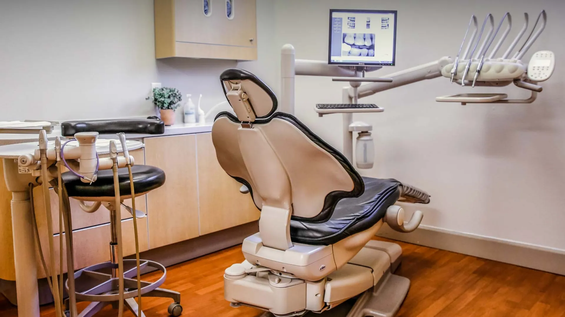
Diagnostics & Technology · 3D Imaging
Large field-of-view CBCT imaging can provide three-dimensional information about teeth, jaws, bone, jaw joints, sinuses, airway anatomy, and surrounding structures when those findings are relevant to dental care.
Traditional dental x-rays remain valuable, but some questions require a broader three-dimensional view. Cone-beam computed tomography (CBCT) can show anatomical relationships that two-dimensional images cannot fully represent.
At Bridgetown Dental, 3D imaging is used selectively, when the additional information can help answer a clinical question, support treatment planning, or identify a finding that may warrant further evaluation.
Why 3D Imaging Matters
The value of CBCT is not simply seeing more anatomy. It is using three-dimensional information selectively when it can clarify a finding, improve planning, or help determine whether another type of evaluation is appropriate.
A large field-of-view CBCT can provide detailed views of teeth, roots, supporting bone, jaw relationships, temporomandibular joints, sinuses, nasal structures, airway anatomy, and other craniofacial structures within the captured field.
An image is one part of an evaluation. CBCT findings are interpreted alongside the patient's history, symptoms, examination, and other appropriate testing. Airway dimensions on a scan, for example, do not by themselves diagnose obstructive sleep apnea or determine how well someone is breathing during sleep.
In selected sleep, airway, TMJ, or orofacial-pain evaluations, 3D anatomy may add context about jaw relationships, joint structures, nasal and sinus anatomy, and the shape of the upper airway. Those findings are considered only as part of the broader clinical picture.
CBCT can also support selected restorative, orthodontic, endodontic, surgical-referral, and implant-related planning by helping clarify anatomy, bone, tooth position, restorative relationships, or other structures that may affect the plan.
More imaging is not automatically better care. The purpose of 3D imaging is to obtain additional information when it is clinically justified and likely to influence evaluation, treatment planning, referral, or coordination with another provider.
From Training to Patient Care
See how 3D imaging can contribute to sleep and airway evaluation when anatomical information is relevant, and how those findings fit alongside symptoms, sleep testing, and physician diagnosis.

We’re here to listen and help you decide what makes sense for you.